Last generation technologies


- PCR – Bio-Rad Laboratories INC CFX Opus 96 DX is an open system that offers maximum flexibility in the rapid development of analyses in a clinical laboratory environment. It can multiplex up to 5 targets per well. The CFX Opus Dx portfolio offers options to manage multiple thruputs and sample volumes. The tools offer precise quantification, accurate thermal cycler performance, and powerful, easy-to-use software.
- Retinograph – Optos Silverstone: it allows a scan at very high speed in order to image the superficial and deep vascular structures both retinal and choroidal. This in turn allows a preoperative evaluation to rule out the presence of occult neovascularizations in patients who will undergo vitreoretinal surgery. The patient’s vascular study can provide information on postoperative functional recovery and support clinical diagnosis in patients with posterior segment oncological diseases
- Mirror Microscope – KONAN CellCheck C Advanced device for corneal endothelial analysis. It uses mirror technology to provide high-resolution images of endothelial cells, facilitating the diagnosis and monitoring of corneal diseases. Its intuitive interface and integrated analysis software allow a quick and accurate evaluation. It is designed to be ergonomic and easy to use, improving efficiency in clinical settings.
- PASCAL Synthesis 577 Retinal Laser allows subthreshold laser treatment, namely a new therapeutic option for patients with diabetic macular edema. The wavelength longer than 577 nm better affects the retinal pigment epithelium with dispersion lower than lasers at 532 or 561 nm.
- RETIMAX ADVANCED PLUS is a sophisticated diagnostic system used in ophthalmology for the evaluation of retinal and electrophysiological function. Equipped with advanced technologies, it allows to perform tests such as ERG (electroretinogram) and VEP (visual evoked potentials) with high precision. The integrated software offers detailed analyses and comprehensive reports, facilitating the diagnosis of retinal and neuro-ophthalmic diseases. Ergonomic design and intuitive interface improve user experience and operational efficiency.
- Mass Spectrometry – Orbitrap Exploris 240 equipped with High-Field Asymmetric-Wavefrom Ion Mobility Spectrometry: it allows an experimental approach of proteomics here applied to ophthalmology in order to explore the pathophysiological bases of ocular diseases, identify biomarkers of diagnostic or prognostic value in biological fluids, develop pharmacological development models and develop diagnostic analysis platforms, mainly in the microbiological field.
- High Performance Liquid Chromatography (HPLC)
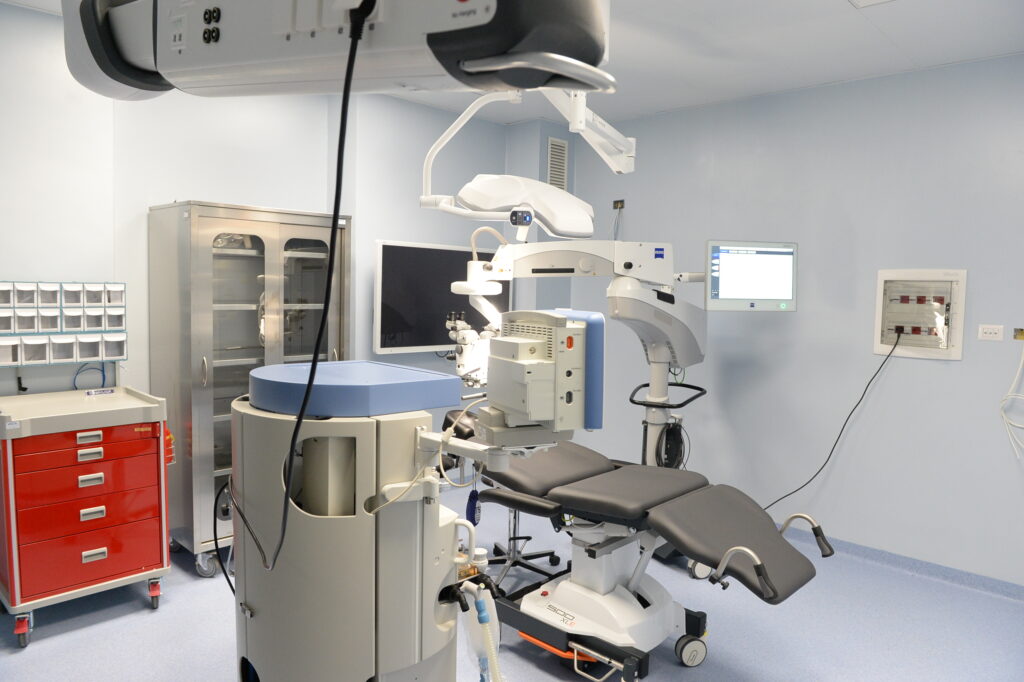
- Leica Microscope Proveo 8: an advanced system designed for eye surgery. It offers superior optical quality thanks to FusionOptics technology, which combines high resolution and extended depth of field. It has an integrated LED lighting for a clear and constant visualization of the eye tissues. Automated autofocus system and digital image control improve accuracy and efficiency during surgeries. The modular configuration of Proveo 8 allows for flexible customization to meet the specific needs of surgeons.
- Octopus 900 Perimeter is an advanced diagnostic tool for visual field assessment. It is equipped with a wide range of standardized and customized tests to detect and monitor diseases such as glaucoma. Integrated EyeSuite technology facilitates data analysis and patient management. The ergonomic design and intuitive interface ensure comfortable and efficient use. The versatility of the Octopus 900 makes it suitable for both clinics and specialist studies.
- Cobra Yag Laser is an avanced medical device used in ophthalmology for the treatment of various ocular pathologies, including posterior capsulotomy and iridotomy. Thanks to its precision, it allows safe and minimally invasive interventions. It features an intuitive interface and ergonomic design, making it easy for professionals to use. High-quality laser technology ensures effective and fast results.
- Angioplex Module leverages OCT angiography (OCTA) technology to visualize retinal microvascularization without the use of dyes. This allows the detailed detection of vascular abnormalities, such as neovascularizations and microaneurysms, improving the diagnosis and monitoring of retinal diseases. The system provides high-resolution images in real time, facilitating timely interventions.